Language


The FluoroScan technique yields qualitative information on the antioxidant capacity of the test substances. This test system is particularly suitable for screening potential new active substances.
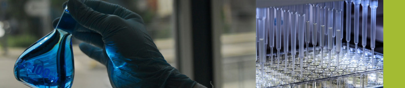
Using a (non-)fluorescent dye, characterised by high sensitivity and ease of use, the intracellular free radical formation is detected. Intracellular free radical formation produces oxidation of the dye, which is then stimulated to fluoresce. Fluorescence is a measure of the quantity of free radicals present. The higher the antioxidant capacity of the cells, the more radicals can be scavenged before the dye is oxidised.
The choice of stressor depends on the question at hand and is adapted to the project requirements. Using multiwell plates, the cells are pre-incubated with the test substances and then subjected to the test procedure. To test for lipophilic test substances, BioTeSys uses several special solubilisation techniques.
Advantages
Supplementary testing
The FluoroScan technique provides preliminary estimations of the antioxidant potential of a test substance quickly and reliably. For a more in-depth evaluation, BioTeSys uses additional cell biology parameters such as cytotoxicity, metabolic activity, proliferation and uptake kinetics.
Metabolic activity
Metabolic activity is measured using the MTT assay detection is based on measuring the turnover activity of mitochondrial dehydrogenase in living cells. The soluble yellow stain (3-[4,5-dimethylthiazol-2-yl]-2,5-diphenyltetrazoliumbromide (MTT)) is converted into violet, insoluble MTT formazan crystals by mitochondrial dehydrogenases. The crystals ca be dissolved again with organic solvents and the violet solution produced can be detected spectrophotometrically.
Approved Quality